Anatomy Through Imaging – Task 1, UD2, CESUR
Anatomy is the cornerstone of medical sciences, providing a detailed understanding of the structure and function of the human body. With advancements in technology, imaging techniques have revolutionized the study of anatomy by allowing non-invasive visualization of internal structures. This article explores various imaging modalities, their applications, advantages, and limitations in the study of human anatomy, specifically for Task 1, Unit 2 (UD2) of the CESUR program.
The Importance of Imaging in Anatomy
Traditional anatomy education relied on cadaver dissection and physical models. While these methods remain essential, modern imaging techniques provide real-time, three-dimensional insights into living bodies, aiding in diagnosis, treatment planning, and medical education. Imaging enhances our ability to study anatomical variations, disease progression, and physiological functions dynamically.
Imaging Modalities in Anatomy
1. X-ray Radiography
Principle: X-ray imaging uses electromagnetic waves to create images based on the varying absorption of radiation by different tissues.
Applications:
- Bone fractures and joint dislocations
- Chest and abdominal evaluations
- Detection of foreign objects
Advantages:
- Quick and widely available
- High resolution for bones
- Cost-effective
Limitations:
- Exposure to ionizing radiation
- Limited soft tissue contrast
2. Computed Tomography (CT)
Principle: CT scanning uses X-rays in a rotating manner to create cross-sectional images of the body, offering detailed views of anatomical structures.
Applications:
- Detailed visualization of bones and soft tissues
- Trauma assessment
- Tumor detection and staging
Advantages:
- High-resolution imaging
- Ability to reconstruct 3D images
- Rapid image acquisition
Limitations:
- Higher radiation exposure
- Expensive compared to X-ray
- Contrast agents may cause allergic reactions
3. Magnetic Resonance Imaging (MRI)
Principle: MRI uses strong magnetic fields and radio waves to generate detailed images of soft tissues without ionizing radiation.
Applications:
- Brain and spinal cord imaging
- Musculoskeletal injuries
- Cardiovascular evaluations
Advantages:
- Excellent soft tissue contrast
- No ionizing radiation
- Multi-planar imaging capabilities
Limitations:
- Expensive and time-consuming
- Contraindicated in patients with metallic implants
- Limited availability
4. Ultrasound (US)
Principle: Ultrasound uses high-frequency sound waves to create real-time images of internal organs.
Applications:
- Obstetrics and fetal imaging
- Cardiac function assessment (echocardiography)
- Abdominal and pelvic evaluations
Advantages:
- No radiation exposure
- Portable and cost-effective
- Real-time imaging
Limitations:
- Limited penetration in obese patients
- Operator-dependent results
- Poor image quality in gas-filled structures
5. Positron Emission Tomography (PET)
Principle: PET imaging detects metabolic activity using radiotracers to identify functional abnormalities in tissues.
Applications:
- Cancer diagnosis and staging
- Neurological disorders (e.g., Alzheimer’s disease)
- Cardiac viability studies
Advantages:
- Functional and metabolic assessment
- Early disease detection
- Combination with CT or MRI enhances accuracy
Limitations:
- High cost and limited availability
- Radiation exposure from tracers
- Time-consuming procedure
Comparative Analysis of Imaging Techniques
| Imaging Modality | Best for | Advantages | Limitations |
|---|---|---|---|
| X-ray | Bones, lungs | Quick, inexpensive | Radiation exposure, limited soft tissue detail |
| CT | Bones, soft tissues, organs | High resolution, 3D reconstruction | High radiation dose, expensive |
| MRI | Brain, muscles, joints | No radiation, excellent soft tissue contrast | Expensive, time-consuming |
| Ultrasound | Fetal, cardiac, abdominal | Safe, portable, real-time imaging | Operator-dependent, limited penetration |
| PET | Cancer, brain function | Functional imaging, early disease detection | Expensive, radiation exposure |
Clinical Applications of Imaging in Different Anatomical Systems
1. Skeletal System
- X-rays and CT scans are essential for detecting fractures, tumors, and joint disorders.
- MRI is preferred for soft tissue injuries such as ligament and cartilage damage.
2. Nervous System
- MRI and CT scans are used for brain and spinal cord pathologies.
- PET scans help in diagnosing neurodegenerative diseases.
3. Cardiovascular System
- Echocardiography (ultrasound) assesses heart function.
- CT angiography and MRI provide detailed images of blood vessels.
4. Respiratory System
- Chest X-rays and CT scans are commonly used for diagnosing lung diseases such as pneumonia and cancer.
5. Gastrointestinal System
- Ultrasound helps detect liver, gallbladder, and intestinal disorders.
- CT and MRI are used for more detailed assessments.
6. Reproductive System
- Ultrasound is the gold standard for fetal imaging and gynecological evaluations.
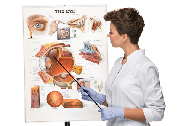
The Role of Imaging in Medical Education
Imaging plays a vital role in anatomy education by allowing students to visualize and analyze anatomical structures dynamically. Digital platforms integrate 3D imaging, enhancing learning and clinical decision-making skills. Virtual reality (VR) and augmented reality (AR) are emerging tools that offer immersive experiences for anatomy education.
Ethical Considerations in Imaging
- Radiation Safety: Minimizing exposure, especially in children and pregnant women.
- Informed Consent: Patients should be aware of risks and benefits.
- Privacy and Confidentiality: Compliance with regulations such as HIPAA and GDPR ensures patient data protection.
Future Trends in Anatomical Imaging
- AI and Machine Learning: Enhancing image interpretation and diagnostic accuracy.
- Hybrid Imaging Techniques: Combining PET-CT or PET-MRI for improved diagnostics.
- Portable and Wearable Imaging Devices: Expanding access to remote areas.
Conclusion
Imaging techniques have revolutionized anatomical studies, offering non-invasive, real-time, and detailed insights into the human body. Each modality has unique strengths and limitations, making their appropriate selection crucial for clinical and educational purposes. As technology advances, the integration of AI, hybrid imaging, and portable solutions will further enhance our ability to diagnose and understand human anatomy effectively.
By understanding the principles, applications, and limitations of each imaging technique, healthcare professionals and students can make informed decisions, improving patient care and medical education outcomes.


